Clinical Image Volume: 1 Issue:1
Keywords
hepatic cyst, IVC compression, pulmonary embolism
Description
Simple hepatic cysts are considered to be a benign disease found in approximately 1-5% of the general population.1 Patients with hepatic cysts infrequently experience complications such as infection, torsion, or spontaneous ruptura.1 Very few cases have documented external compression of nearby structures by hepatic cysts, leading to inferior vena cava (IVC) or right atrial compression.1-3 Compression of the IVC (Figure 1A) can lead to venous stasis and subsequent thrombus formation (e.g. deep vein thrombosis, pulmonary embolism).
Contrast-enhanced CT imaging can be used to confirm the presence of a large simple cyst exerting mass effect on the IVC (Figure 1A). One potential treatment option involves percutaneous drainage of the cyst for decompression (Figure 1B).1 For more advanced cases, laparoscopy, mechanical thrombectomy, and anti-thrombolytic therapy may be warranted.1,2 Additional testing may be indicated to rule out conditions such as malignancy, echinococcus infection, Entamoeba histolytica infection, or autosomal dominant polycystic kidney disease.
Following reduction or resolution of extrinsic compression, IVC filter placement and oral anticoagulation is often used.2,3 This case showcases a rare complication of hepatic cysts and exemplifies the importance of close follow-up and drainage.
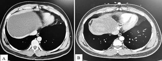
Figure 1 Computed Tomography (CT) imaging with contrast of the abdomen of a 38 year old male demonstrating a large hepatic cyst before (A) and after
(B) percutaneous drainage. Prior to intervention, the hepatic cyst caused extrinsic compression of the inferior vena cava (IVC) (A), contributing to the development of bilateral pulmonary emboli. He had no risk prior risk factors for thrombosis. In Figure 1B, after placement of an IVC filter and deflation of the cyst via percutaneous drainage, the hepatic cyst is significantly decreased in size with resolution of IVC compression.
Acknowledgements
None.
Conflict of interest
The authors declare that they have no competing interests.
References
- Nakabayashi K, Murakami M, Hata S, et al. Giant Hepatic Cyst: A Possible Cause of Inferior Vena Cava Syndrome. Intern Med. 2021;60(13):2081–2084.
- Musielak M, Singh R, Hartman E, et Simple Hepatic Cyst Causing Inferior Vena Cava Thrombus. Int J Surg Case Rep. 2014;5(6):339–341.
- Ko M, Kim T, Lee W, et al. Deep Vein Thrombosis Due to Compression of Huge Hepatic Cyst Successfully Treated by Inferior Vena Cava Filter and Cyst Drainage. Korean J Gastroenterol. 2018;72(3):146–149.