Case Report Volume: 1 Issue:1
Abstract
Background: Acute appendicitis during pregnancy is a rare but urgent surgical condition that can have serious maternal and fetal consequences if not promptly managed. Diagnosis is often complicated by physiological and anatomical changes in pregnancy.
Case presentation: We present the case of a 33-year-old woman at 33 weeks and 4 days of gestation who was admitted with acute right lower quadrant abdominal pain, associated with nausea and localized tenderness. Clinical examination and abdominal ultrasound suggested acute appendicitis. An appendectomy was performed without intraoperative complications. Postoperative recovery was uneventful, and both maternal and fetal conditions remained stable throughout the hospital stay.
Conclusion: This case underscores the importance of maintaining a high index of suspicion for acute appendicitis in pregnant patients presenting with abdominal pain. Prompt surgical intervention via laparotomy, particularly in advanced gestation, can result in favorable outcomes for both mother and fetus.
Introduction
Acute appendicitis is the most frequent non-obstetric surgical indication in pregnant women, nevertheless, it remains rare with an incidence of 1/ 500 to 1/635.1 Acute appendicitis occurs most often during the second trimester of pregnancy 45% due to the anatomical changes that happen during this period,2 the appendix is pushed upwards as the fetus grows and the uterus enlarges. The clinical diagnosis of acute appendicitis in pregnant women is often mistaken for another etiology. In this case, we find guarding and pain on sudden decompression of the right iliac fossa but pain at the MacBurney point may be absent. Complementary examinations are necessary in order to make the diagnosis of acute appendicitis, mainly ultrasound and biological workup.3 The curative treatment of acute appendicitis remains appendectomy, either by laparoscopy or by laparotomy, which obviously has fewer advantages than the former, but which has its place in our context. And for this, prevention of a premature delivery is required. Acute appendicitis in pregnant women is a major problem requiring cooperation between different specialties to provide better maternal-fetal care.
Case report
We hereby present the uncommon case of a 33 years old paucigravidae patient with no medical or surgical pathological history, current pregnancy estimated at 33 weeks and 4 days admitted to the emergency department complaining of acute continuous right lower quadrant abdominal pain gradually intensifying for the last 3 hours and bilious vomiting for the last 24hours. Her obstetric follow up was uneventful. She had no history of abdominal trauma or digestive disease. Her general examination and physical assessment revealed abnormal vitals. The patient was febrile at 38.1 Celsius, her blood pressure was normal 110\70 mmHg, and her pulse was high at 120 bpm with 99 oxygen saturation at room air, there was no jaundice. Abdominal examination shows a relaxed gravid uterus with a uterine height of 28 cm corresponding to 33 weeks and a right hypochondrium guarding. For the gynecological examination, the cervix was closed with no vaginal discharge or amniotic fluid. These clinical findings allowed the suspicion of acute appendicitis.
Her cellular blood count showed a normal hemoglobin at 12g\dl with elevated blood count at a rate of 21600 \ul. Her platelets and PT were within normal range. C reactive protein was high up to 132,70. An abdomino pelvic ultrasound was performed not being conclusive due to the abnormal position of the appendix hidden by the gravid uterus, a CT-scan was then done revealing an enlarged swollen and undifferentiated latero caecal appendice measuring 13mm, appendiceal wall thickening with a peri appendiceal fat infiltration and some infracentimetrique ganglions of which the largest measures 7mm.
The surgical treatment performed by the visceral surgeon was an open appendectomy, an incision at the point of McBurney was made, at the exploration there is no effusion or pus, the appendix was of backseat and under serous, a retrograde dissection of the appendix after section of its base and ligation section of the mesoappendicuary, the appendicular base was healthy and was closed by a denier point (Figure 1). The operative suites were simple, and output at day 3.
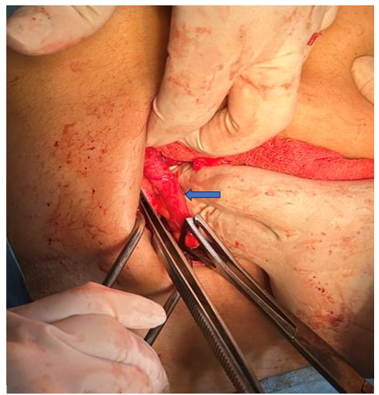
Figure 1Intraoperative finding. Blue arrow: Acute Suppurative Appendicitis
The parturient did not benefit from tocolysis and did not present a threat of premature delivery. Her pregnancy continued naturally. She went into spontaneous labor at 39 weeks, resulting in vaginal delivery of a healthy female newborn.
Discussion
The incidence of a digestive emergency is estimated to be 1/500 pregnancies, half of which require surgical management.4 Acute appendicitis is common in young adults. Although its occurrence during pregnancy is rare (1/2000 pregnancies), it is the most frequent non-obstetric surgical emergency (25%) during pregnancy.4,5 The similarities between the signs of AA and the sympathetic signs of pregnancy are a source of misdiagnosis and therapeutic delay, which contribute to its seriousness.6 Acute appendicitis is the most frequent non-traumatic digestive emergency during pregnancy.7,8 The prevalence of the condition during pregnancy is in the order of 0.05-0.1% in Western series.7 Lebeau et al.,6 reported a prevalence of 0.2%. The prevalence of the disease is highest in the first two trimesters of pregnancy.2 The incidence per trimester is 32, 42 and 26%.2 Marret et al.,8 and Chambon & Quandalle9 reported frequencies of 33.3% and 66.6% respectively. The prevalence of the condition during pregnancy is low, ranging from 0.05% to 0.1% in Western series. In the series by Nouira et al.,10 it was described that the first two trimesters of pregnancy were the most affected. This was not the case for Halvorsen et al.,11 who reported a series of 12 appendectomies, with equal frequency in the 2nd and 3rd trimesters of pregnancy. Gestational age therefore appears to have no influence on the occurrence of acute appendicitis.
The diagnosis of acute appendicitis is more difficult during pregnancy. Some clinical signs are mistakenly attributed to the sympathetic signs of pregnancy. Pregnancy brings with it a number of anatomical and physiological changes, which may explain the difficulty and sometimes delay in diagnosis. Late diagnosis of appendicitis can lead to serious maternal and fetal complications.12,13 Classically, the diagnosis of AA outside pregnancy is evoked by the association of pain in the right lower quadrant, nausea or vomiting, fever and tenderness on palpation of the right lower quadrant. In the event of pregnancy, these signs may be altered or unrecognized, resulting in severe forms of the disease due to delayed diagnosis. Clinically, the location of abdominal pain leads to a prioritization of other diagnoses over acute appendicitis. The unusual location of pain in appendicular inflammation in the right flank (FD) and right hypochondrium (HCD) leads practitioners to first evoke acute pyelonephritis, which is more frequent during pregnancy.14 This is a source of diagnostic error. Moreover, clinical signs in the first trimester, when the uterus is not enlarged, are no different from those observed in non-pregnant women. These include pain in the right iliac fossa, often associated with fever, which may lead to discussion of a threatened early abortion or urinary tract infection, hence the importance of obstetrical examination and cytobacxteriological urine exam. Nausea and vomiting are common at this stage of pregnancy, and are the first sympathetic signs of pregnancy, not usually a cause for concern. Although gravidic vomiting is commonplace at this stage of pregnancy, it should motivate careful palpation of the abdomen, especially as the symptoms of acute appendicitis may be incoercible.
Abdomino-pelvic ultrasound confirms the diagnosis when it visualizes an incompressible appendix over 7 mm in diameter, aperistaltic with a parietal thickness of over 3 mm and sometimes the presence of fluid in the appendicular lumen.15 Performed by a trained operator, ultrasound has a sensitivity of 100% and a specificity of 96% at this gestational age.16 It can reveal indirect signs such as a small, free effusion in the right iliac fossa or in the cul de sac of Douglas. It can confirm the diagnosis of acute appendicitis in 40% of cases.17,18 It is also useful for ruling out associated adnexal or obstetric pathology, and for documenting pregnancy by specifying gestational age and fetal vitality. The blood count is difficult to interpret due to the physiological hyperleukocytosis of pregnancy,10 while the CRP may be normal. These two tests are therefore of little value in diagnosing appendicitis during pregnancy.
During the last two trimesters of pregnancy, the diagnosis of appendicitis becomes more difficult due to a change in the anatomical location of the appendix and the uterus: ascension of the appendix by the volume taken on by the gravid uterus, which becomes twenty times larger.19 The appendix is pushed up and out, reaching the costal margin by the 8th month.8 Pain is localized in the right flank or hyponchondrium. It may be accompanied by uterine contractions, suggesting a threat of late abortion or premature delivery. A fever is observed in 50% of cases.7-12 Nausea and vomiting are observed in 87% of cases.13 Ultrasound may be hampered in the last two trimesters by uterine volume. The upwardly displaced appendix is less accessible to ultrasound in the subhepatic position than in the right iliac fossa. However, ultrasound can reveal indirect signs such as a sub-diaphragmatic effusion; in advanced forms, it can easily find a peritoneal effusion, as in the first trimester; it is also of interest in the differential diagnosis with other causes of abdominal pain during pregnancy. In our case the ultrasound failed to make a diagnosis of the acute appendicitis, so a contrast-enhanced CT scan was performed revealing an enlarged, non-compressible appendix, with wall thickening, peri appendiceal fat stranding, and mild free fluid-findings consistent with acute appendicitis.
In the event of abdominal pain, the first thing to do is to rule out obstetrical emergencies, which could jeopardize the mother's vital prognosis: retroplacental hematoma or uterine rupture. Right-sided urinary tract infections are frequent at this gestational age, due to the dextrorotation of the uterus, which compresses the right ureter, and the reduction in ureteral peristalsis caused by gravid hormonal impregnation.13 These two phenomena contribute to urinary stasis, leading to frequent bacterial proliferation in the urine. The complications of acute appendicitis during pregnancy are the same as outside pregnancy. In the 1st trimester, all the complications of appendicitis can be seen. At this time, the uterus, still pelvic, does not displace the surrounding organs, which may form adhesions around the appendicular focus and isolate it from the peritoneal cavity, creating an appendicular plastron. Appendicular crises can also evolve into acute diffuse peritonitis. In the last two trimesters, there is no reason why appendicular infection should not become widespread. Uterine contractions hinder the formation of adhesions and the compartmentalization of the infection; high steroid levels reduce the inflammatory response: and increased pelvic vascularization facilitates the spread of infection. For all these reasons, peritonitis develops more rapidly in the 3rd trimester.
The treatment of acute appendicitis in pregnant women is surgical. It consists of an appendectomy. The choice of approach depends on several factors, including gestational age, the stage of progression of the appendicitis, the patient's build, the presence of abdominal scarring and the surgeon's preference. A Mac Burney incision, enlarged, if necessary, enables appendectomy to be performed easily in the first trimester. In the last two trimesters, the incision should be higher, in the right flank, as performed in our case. These high incisions resolve the operative difficulties associated with cecal migration in the second and third trimesters. For some authors, the caeco-appendicular region would be better exposed by a median subumbilical incision in the second trimester, and by a Jalaguier incision in the second and third trimesters.6 In the case of diffuse appendicular peritonitis, a median incision straddling the umbilicus allows rapid access and meticulous exploration of the abdominal cavity.
Acute appendicitis outside pregnancy is a documented and validated indication for a laparoscopic approach.20 In pregnancy, this approach raises many fears: damaging the gravid uterus, injuring it during trocar insertion and altering fetal well-being through CO2 pneumoperitoneum.21,22 However, the literature reports new series of laparoscopies carried out even after 28SA, with favorable outcomes for both fetus and mother.23 Sadot et al.,24 reported a series of 65 pregnant patients operated on with the diagnosis of acute appendicitis. Forty-eight patients underwent laparoscopic surgery. The authors concluded that this approach was feasible and safe in all trimesters of pregnancy. The benefits of this less invasive surgery in terms of reduced parietal effraction, reduced postoperative pain, faster resumption of transit and early lifting seem to be quite decisive and valuable assets during pregnancy. Laparoscopic surgery reduces the risk of thromboembolism through rapid rehabilitation and less postoperative pain.25 However, this risk seems to be increased by pregnancy and pneumoperitoneum.26 For all its advantages, several authors are in favor of widespread use of laparoscopy during pregnancy.23,27,28 Technically, the patient is positioned in dorsal decubitus during the first trimester, with a 20 to 30◦ roll to the left, enabling a left lateral decubitus to be reproduced from 15 SA onwards.24 The pneumoperitoneum can be created using either the open laparoscopic technique or the Palmer needle, depending on the operator's choice. The site of insertion of the needle or optical trocar also depends on the surgeon's choice, but also on gestational age and uterine volume (umbilicus, supraumbilical region, left hypochondrium). The uterine fundus must be located by palpation and/or ultrasound. CO2 pressure should be kept below 12 mmHg.22,24 It is conventional to limit the number of trocars to three. In the first trimester, the rule of triangulation is respected (the median trocar is inserted higher up than the other trocars); but during the last two trimesters, the trocars may be inserted more grouped together and on one side of the gravid uterus. Intraoperatively, it is essential to rapidly treat any maternal hypotension secondary to anesthesia, to limit uteroplacental hypoperfusion and subsequent fetal suffering. Certain devices, such as the Trendelenburg position, can be used to facilitate laparoscopic access to the appendix. Mobilization of the uterus should only be carried out indirectly, by mobilizing the utero-ovarian ligament.
The prescription of thromboprophylaxis is debatable, depending on the patient's history, maternal weight, term of pregnancy, the procedure to be performed and the duration of pneumoperitoneum in the case of laparoscopic approach.25 Postoperative tocolysis remains a controversial issue. Some authors recommend it systematically.6,10,11,25 Pearl et al.,28 consider that systematic preventive tocolysis has no place and is only recommended in cases of threatened preterm delivery. In our case the patient received 5 days of thromboprophylaxis and no tocolysis was prescribed.
Maternal-fetal prognosis depends on the severity of the condition and the delay in treatment. The fetal death observed in a case of appendicular perforation peritonitis diagnosed and treated 4 days after admission of the pregnant woman is a case in point. Fetal mortality rises to over 35% in cases of appendicular peritonitis29 and varies between 1 and 8% in women with acute uncomplicated appendicitis.29,30 We noted one premature delivery that was unrelated to appendicular disease. Prematurity and spontaneous delivery were the main fetal risks. The prematurity rate was 22.2%. In the series by Nouira et al.10 These risks are particularly high during the first week after appendectomy. No maternal deaths were noted in our work, as in other series.5,10,11 This is due to early diagnosis and treatment. In our case, the diagnosis was made before 24 hours of admission and the pregnant woman was operated on immediately.
Conclusion
Acute appendicitis in pregnant women is a rare condition, and a surgical emergency that can be life-threatening for the mother and fetus. Diagnosis is difficult and requires rigorous analysis of clinical and paraclinical data. In the 1st trimester, diagnosis is straightforward, treatment simple and prognosis generally good. In the last 2 trimesters, diagnostic difficulties are responsible for severe forms and aggressive surgical treatment. Pelvic ultrasound and cytobacteriological examination of urine should be systematically performed in the event of abdominal pain in pregnant women. Treatment is surgical, involving appendectomy. Morbidity and mortality are not negligible. This case report has been reported in line with the SCARE Criteria.31
Ethical approval
Ethics approval has been obtained to proceed with the current study.
Consent approval
Written informed consent was obtained from the patient for publication of this case report and any accompanying images. A copy of the written consent is available for review by the Editor-in-Chief of this journal.
Acknowledgements
The authors thank all the participants for their efforts in responding to the scales as well as the editors of this periodical for their constructive comments.
Conflict of interest
The authors declare that they have no competing interests.
References
- Franca Neto AHD, Amorim MMRD, Nóbrega BMSV. Acute appendicitis in pregnancy: literature review. Rev Assoc Médica Bras. 2015;61(2):170–177.
- Borst AR. Acute appendicitis: Pregnancy complicates this diagnosis. J Am Acad Physician Assist. 2007;20(12):36–41.
- Terasawa T, Blackmore CC, Bent S, et al. Systematic Review: Computed Tomography and Ultrasonography To Detect Acute Appendicitis in Adults and Adolescents. Ann Intern Med. 2004;141(7):537–546.
- Germain A, Brunaud L. Chirurgie viscérale et grossesse. J Chir Viscérale. 2010;147(3):182–189.
- Allen JR, Helling TS, Langenfeld M. Intraabdominal surgery during pregnancy. Am J Surg. 1989;158(6):567–569.
- Lebeau R, Diané B, Koffi E, et al. Appendicite aiguë et grossesse: À propos de 21 cas. J Gynécologie Obstétrique Biol Reprod. 2005;34(6):600–605.
- Weston AR, Jackson TJ, Blamey S. Diagnosis of appendicitis in adults by ultrasonography or computed tomography: A systematic review and meta-analysis. Int J Technol Assess Health Care. 2005;21(3):368–379.
- Henri M, Jacques L, Marc L, et al. Urgences chirurgicales au cours de la grossesse. Obstétrique; 2000.
- Chambon JP, Quandalle P, Régnier C, et al. Non-gynecological abdominal emergencies during pregnancy. Ann Chir. 1986;40(7):455–461.
- Nouira M, Jerbi M, Sahraoui W, et al. Acute appendicitis during pregnancy: A review of 18 cases.
- Halvorsen AC, Brandt B, Andreasen JJ. Acute appendicitis in pregnancy: complications and subsequent management. Eur J Surg. 1992;158(11–12):603–606.
- Murariu D, Tatsuno B, Hirai CAM, et al. Case report and management of suspected acute appendicitis in pregnancy. Hawaii Med J. 2011;70(2):30–32.
- Basaran A, Basaran M. Diagnosis of Acute Appendicitis During Pregnancy: A Systematic Review. Obstet Gynecol Surv. 2009;64(7):481–488.
- Fournié A, Jalle T, Sentilhes L, et al. Infections urinaires chez la femme enceinte. EMC-Obstétrique. 2008;3(3):1–8.
- Taourel P, Kessler N, Blayac PM, et al. Imagerie de l’appendicite: échographie, scanner ou rien du tout? J Radiol. 2002;1132(2502):203.
- Lim HK, Bae SH, Seo GS. Diagnosis of acute appendicitis in pregnant women: value of sonography. Am J Roentgenol. 1992;159(3):539–542.
- Bretagnol F, Zappa M, Panis Y. Place de l’imagerie dans le diagnostic d’appendicite aiguë. J Chir (Paris). 2009;146(5):8–11.
- Douglas CD. Randomised controlled trial of ultrasonography in diagnosis of acute appendicitis, incorporating the Alvarado score. BMJ. 2000;321(7266):919–922.
- Hodjati H, Kazerooni T. Location of the appendix in the gravid patient: a re-evaluation of the established concept. Int J Gynecol Obstet. 2003;81(3):245–247.
- Pirro N, Berdah SV. Appendicites: cœlioscopie ou non? J Chir. 2006;143(3):155–159.
- Lyass S, Pikarsky A, Eisenberg VH, et al. Is laparoscopic appendectomy safe in pregnant women? Surg Endosc. 2001;15(4):377–379.
- Palanivelu C, Rangarajan M, Parthasarathi R. Laparoscopic appendectomy in pregnancy: a case series of seven patients. JSLS. 2006;10(3):321–325.
- Barnes SL, Shane MD, Schoemann MB, et al. Laparoscopic appendectomy after 30 weeks pregnancy: report of two cases and description of technique. Am Surg. 2004;70(8):733–736.
- Sadot E, Telem DA, Arora M, et al. Laparoscopy: a safe approach to appendicitis during pregnancy. Surg Endosc. 2010;24(2):383–389.
- Colomb S, Bonnin M, Bolandard F, et al. Prise en charge anesthésique de la femme enceinte pour cœliochirurgie gynécologique non obstétricale à la maternité de Clermont-Ferrand. Ann Fr Anesth Réanimation. 2006;25(1):11–16.
- Holzheimer RG. Laparoscopic procedures as a risk factor of deep venous thrombosis, superficial ascending thrombophlebitis and pulmonary embolism-case report and review of the literature. Eur J Med Res. 2004;9(9):417–422.
- Bisharah M, Tulandi T. Laparoscopic surgery in pregnancy. Clin Obstet Gynecol. 2003;46(1):92–97.
- Pearl J, Price R, Richardson W, et al. Guidelines for diagnosis, treatment, and use of laparoscopy for surgical problems during pregnancy. Surg Endosc. 2011;25(11):3479–3492.
- Babaknia A, Parsa H, Woodruff JD. Appendicitis during pregnancy. Obstet Gynecol. 1977;50(1):40–44.
- Mahmoodian S. Appendicitis complicating pregnancy. South Med J. 1992;85(1):19–24.
- Sohrabi C, Mathew G, Maria N, et al. The SCARE 2023 guideline: updating consensus Surgical CAse REport (SCARE) guidelines. Int J Surg. 2023;109(5):1136–1140.